Affiliated Dermatologists provides comprehensive, state-of-the-art treatment for issues relating to the skin, hair, and nails. With years of experience practicing, teaching, and researching the cutting edge of dermatology, Affiliated Dermatologists is experienced in determining the treatments and products that will truly optimize the appearance and health of skin at any age.
To provide you the most up-to-date dermatological care, we perform the latest procedures, including:
Cryosurgery
Cryosurgery (cryotherapy) is the application of extreme cold to destroy abnormal or diseased tissue. A common method of freezing lesions is to use liquid nitrogen as the cooling solution. This super-cooled liquid may be sprayed on the diseased tissue, circulated through a tube called a cryoprobe, or simply dabbed on with a cotton or foam swab. The tissue is destroyed by this application of intense cold. It is rapidly frozen with liquid nitrogen and then slowly thawed. A second freeze is often done to ensure adequate tissue destruction.
Upon treatment, patients experience minor-to-moderate localized pain and redness. Sometimes blisters may form, but these usually scab over and peel away within several days. The area heals with healthy skin. Minor discoloration or scarring is possible with these treatments.
Cutaneous Surgery
Many forms of cutaneous surgery are performed at Affiliated Dermatologists. Simple shave or punch removal of moles to complex removal of birthmarks and all forms of skin cancer. Occasionally electrodessication and curettage may be appropriate. These procedures are performed on the office under local anesthesia. Larger tumors may need staged (multiple) surgeries or specialized closures such as flaps or grafts. Laser surgery is usually used for treatment of scars, birthmarks and anti-aging.
Hand and Foot NBUVB
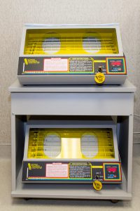 Offered in the convenience of our Brookfield office, this light therapy is effective for psoriasis and eczema of the hands and feet.
NBUVB Narrowband Ultraviolet B Light. The hands and feet are then placed in a special box that produces narrowband ultraviolet B light.
Treatments are typically administered two to three times a week. It will often take 2 months for your condition to improve. Appointment times are flexible and are offered before and after work or school.
Mohs Surgery
MOHS MICROGRAPHIC SURGERY DEFINED
Mohs micrographic surgery involves surgical excision of cancer-containing tissues and systematic microscopic examination of all cut surfaces. It is a highly specialized procedure for the total removal of skin cancers.
Unlike most surgical cancer removals, Mohs uses frozen sections stained with dyes to help identify different skin edges. The freezing process allows for an immediate examination.
MOHS SURGERY STEP BY STEP
This method involves five separate steps:
- Removal of the bulk of the cancer with a skin scraper (a curette).
- Surgical removal of a thin layer of skin.
- Drawing a map and preparing stained frozen tissue sections.
- Examination of the excised tissue under the microscope. After the tissue is removed, it is marked with colored dyes to distinguish the different skin edges.
- The entire procedure (except step 1) is repeated but only in the area of the remaining cancer. Only by careful, systematic microscopic examination of the removed skin can one be as certain as possible that no cancer remains.
During Mohs micrographic surgery, the tissue is examined in a different and more thorough manner than is normally performed by a pathologist associated with an operating room. Mohs micrographic surgery examines the entire sides and undersurface of the excised tissue. If one looks at a loaf of bread, Mohs micrographic surgery examines the whole crust rather than a few slices of the loaf.
FREQUENTLY ASKED QUESTIONS
How long does Mohs micrographic surgery take?
Total removal of a skin cancer, which may involve several surgical stages, is usually completed in one day. After the surgery, a decision is made as to the best way to manage the wound created by the surgery.
How effective is Mohs micrographic surgery?
Using the Mohs micrographic surgical technique, the percentage of success is very high, often 95% to 99%, even if other forms of treatment have failed. Therefore, with this technique, an excellent chance of cure is achieved. However, no one can guarantee a 100% chance of cure.
Will the surgery leave a scar?
Yes. Most forms of therapy will leave a scar. However, the Mohs micrographic surgical procedure tends to minimize this as much as possible. After the wound is healed, you may wish to have the scar improved. Generally, time alone will improve all scars.
What are the advantages of Mohs micrographic surgery?
After the initial tissue is removed, the surgeon can pinpoint with the microscope the areas where there is cancer and then selectively remove tissue only from those areas in the following surgical stages. In this way, the skin cancer is traced out to its roots with little guesswork involved, which results in:
- The removal of as little normal tissue as possible.
- The highest chance of curing the patient (under certain circumstances).
What are the disadvantages?
Mohs micrographic surgery is a process that may involve several surgical stages. The time for this procedure may take several hours and often all day.
What’s the next step after Mohs micrographic surgery?
When we have determined that the skin cancer has been completely removed, a decision is made about what to do with the wound created by the surgery. Usually, there are two choices:
- To let the wound heal by itself (granulation).
- To repair (close) the wound with stitches (either by bringing the wound edges together, or with a skin flap or skin graft).
What happens if I do not have my skin cancer treated?
All types of skin cancer will grow and invade nearby tissue. How fast a skin cancer will grow is unpredictable and varies from person to person. Sometimes skin cancer will destroy important structures such as the nose, lip or eye. Occasionally, skin cancers can be life threatening.
UNDERSTANDING SKIN CANCER
Cancer is tissue which grows at an uncontrollable and unpredictable rate. In the skin, there are three main forms: basal cell carcinoma, squamous cell carcinoma and malignant melanoma. The names refer to the cell types in the top skin layer (the epidermis) from which these cancers are derived.
The most common types of skin cancers are basal cell carcinoma and squamous cell carcinoma. If not removed completely, basal cell carcinoma and squamous cell carcinoma can enlarge from the point where they first occur, and can invade and destroy structures in their path. Basal cell carcinoma is unlikely to spread to distant parts of the body (metastasize). However, some squamous cell carcinomas can metastasize. These types of skin cancers are generally recognized in their early stages and are therefore easily cured.
Malignant melanoma, on the other hand, may be life threatening if not treated early. It usually appears as a brownish-black spot or bump on the skin which enlarges and sometimes bleeds. Sometimes melanomas arise in moles which have been present for many years.
Narrow Band UVB
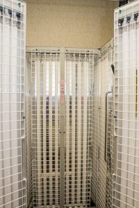 Narrow Band UVB has proven to be an effective and safe treatment psoriasis, eczema, vitiligo, and mycosis fungoides.
We offer this service in the convenice of our Brookfield office. Treatments are typically administered two to three times a week. It will often take 2 months for your condition to improve. Appointment times are flexible and are offered before and after work or school.
Why Narrow Band UVB is different
Conventional broad band UVB lamps emit a variety of wavelengths ranging from 280-330 nm. Narrow Band UVB virtually eliminates superfluous and harmful UV by emitting only wavelengths 311-312 nm. Clinical studies show the peak therapeutic effectiveness of UVB to be within the range of 295-313 nm, but wavelengths below 300 nm can cause erythema or severe burning and increase the risk of skin cancer.
Benefits of Narrow Band Phototherapy
Eliminating UV in wavelengths below 311 nm permits higher intensities and longer exposure times, so patients can derive the maximum benefit from phototherapy. This increased effectiveness permits more aggressive treatment regimens, resulting in a shorter course of treatment.
Patch Testing
Patch testing is a common diagnostic tool used to identify the specific agent that triggers contact dermatitis, an itchy rash that can breakout on your skin in response to a substance to which you are allergic. Common trigger substances include poison ivy, jewelry, hair care products, cleaning solution, detergent, cosmetics, perfumes, and latex rubber.
Most dermatologists use the TRUE Test, which is a very good screening tool but is limited to testing 28 chemicals. At Affiliated Dermatologists we offer an extensive patch testing. We have access to 100’s of allergens which can be special ordered for you if necessary.
The patch testing procedure is simple and painless. In order to identify specific triggers, several patches will be taped onto the skin on the back containing various potential chemicals. These chemicals are then left on the skin for 48 hours, and must remain dry during this time. After 48 hours, the patch test is removed and an initial reading is taken to observe any reactions. An additional reading may be taken after another 24-48 hours. Before undergoing patch testing, patients should stop using oral and topical corticosteroids in the area to be tested, avoid oral antihistamines, and should not expose the test area to the sun for at least three weeks.
When reading the results, the doctor may classify each spot on a scale from negative (meaning no reaction) to extreme reaction (meaning positive results for substance). Strong results may cause blisters or ulcers on the skin, which can be treated once the test is complete.
Once positive results have been determined, patients can take steps to avoid their triggers and prevent contact dermatitis from occurring. We provide a comprehensive list of products a patient may use based on their individual test results and specific information on your individual triggers and how to prevent contact.
Photodynamic Therapy (PDT)
Photodynamic therapy, or Blue Light, is a treatment that uses special drugs, called photosensitizing agents, along with light to kill targeted cells. The procedure can effectively treat early changes in the skin that may lead to skin cancer.
A natural chemical Aminolevulinic acid (Levulan® Kerastick) is a solution that is applied directly to the lesions or spots on the face or scalp to treat actinic keratosis lesions, and causes the lesions to become sensitive to the light. At a prescribed interval after the drug is applied, the nurse will expose the area being treated to a blue light for a period of minutes determined by the doctor. During the light therapy you will wear protective eyewear. You may feel stinging or burning once the area is exposed to the blue light, but it should go away within a day or so. The treated area may get red and scale and crust for up to2-4 weeks before healing.
To achieve maximum improvement of pre-cancerous sun damaged skin, a series of treatments 2 to 4 weeks apart is most effective. After a series of treatments have been completed, treatments can be done at periodic intervals in the future to maintain the rejuvenated appearance of the skin.
Skin Biopsy
During a skin biopsy, we remove a small piece of skin, and send it for evaluation under the microscope. This procedure is performed in order to diagnose, and not to treat the condition. Using the information from the biopsy, the cells under the microscope, we can help treat the condition because we can understand it better.
As the patient, you will experience a small “Prick” from the needle, which delivers a small amount of local anesthetic into the lesion. After that point, you will not feel any pain. A small piece of skin is then removed for evaluation. The risks to this procedure include bleeding, scarring and possible infection. We are equipped to handle any complications or diagnosis arising from the biopsy.


